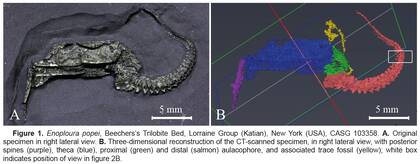
CT scans of Enoploura popei (Echinodermata: Stylophora: Anomalocystitidae) – Supplementary material to Boisset, T. et al. (2024): "Insights into stylophoran anatomy and taphonomy based on an exceptionally preserved mitrate from the Lorraine Group (Upper Ordovician) of New York, USA"
| DOI: | https://doi.org/10.57756/ammjqp |
|---|---|
| Creator(s): |
|
| Publication date: | 13 Jan 2025 |
| Publisher: | Naturhistorisches Museum Wien (NHMW) |
| Collections: | Central Research Laboratories , Geology & Palaeontology |
| Instrument: | YXLON FF35 CT |
| Resource type: | Dataset |
| Specimen: | CASG 103358 |
| License: |
CC BY 4.0 International |
| Tags: | Microct Palaeontology Soft-tissue preservation Stylophora |
| Countries: |
|
| Chronology: | Katian |
| Taxa: | Echinodermata |
| Share: |
|
Abstract
MicroCT scans of pyritized specimen of the anomalocystitid mitrate Enoploura popei Caster, 1952 (Echinodermata: Stylophora: Anomalocystitidae) from Beecher’s Trilobite Bed (Lorraine Group, Katian, Upper Ordovician) in upstate New York (USA) with soft tissue preservation providing insight on the internal anatomy and taphonomy of stylophorans.
Usage Notes
There are two microCT scans available for this specimen:
MicroCT Scan (NHMW)
System: YXLON FF 35 CT
Beam: FXE Transmission Beam
Detector: Perkin Elmer Y.Panel 4343 CT
Scan Parameters: 130 kV, 220 µA, 2000 ms exposure time, 4288 projection images, helical scan
Isotropic voxel size: 9.6 µm
Converted to TIF in Dragonfly software.
MicroCT Scan (MATEIS lab, INSA)
System: Phoenix v|tome|x
Scan Parameters: 140 kV, 80 µA, 667 ms exposure time, 1200 projection images
Isotropic voxel size: 20 µm
Publications
Boisset, T., Lefebvre, B., Mooi, R., Kroh, A., Winkler, V., Adrien, J. & Martin, M.J. (2024) Insights into stylophoran anatomy and taphonomy based on an exceptionally preserved mitrate from the Lorraine Group (Upper Ordovician) of New York, USA. Cahiers de Biologie Marine, 65, 511-516.
Downloads
| File | Description | Size |
|---|---|---|
| CASG-103358-Enoploura-popei_Scan_INSA_20mic.zip | Volume file (.VOL and metadata files) of the microCT scan performed at INSA | 1293.93 MB |
| CASG-103358-Enoploura-popei_Scan_INSA_20mic_TIF.zip | Reconstructed TIF image stack and metadata files of the microCT scan performed at INSA | 1266.42 MB |
| CASG-103358-Enoploura-popei_Scan_NHMW_9-6mic_ReconInfos.xml | Reconstruction parameters (.XML) of the microCT scan performed at the NHMW | 0.01 MB |
| CASG-103358-Enoploura-popei_Scan_NHMW_9-6mic_ScanInfos.xml | Scanning parameters (.XML) of the microCT scan performed at the NHMW | 0.01 MB |
| CASG-103358-Enoploura-popei_Scan_NHMW_9-6mic_TIF.zip | Reconstructed TIF image stack of the microCT scan performed at the NHMW | 9236.08 MB |

 https://orcid.org/0000-0002-3803-9176